Cardiac Image Analysis
(a) Cardiac MR cine image sequence.
(b) Probabilistic label map estimate.
(c) Segmentation of the myocardium.
Publications:
- W.Bai, W. Shi, A. de Marvao, T.J.W. Dawes, D.P. O’Regan, S.A. Cook, D. Rueckert. A bi-ventricular cardiac atlas built from 1000+ high resolution MR images of healthy subjects and an analysis of shape and motion. Medical Image Analysis, 26(1):133-145, 2015. [data]
- W Bai, W Shi, DP O’Regan, T Tong, H Wang, S Jamil-Copley, NS Peters and D Rueckert. A probabilistic patch-based label fusion model for multi-atlas segmentation with registration refinement: Application to cardiac MR images. IEEE Transactions on Medical Imaging, 32(7):1302-1315, 2013. [source code]
- W. Bai, W. Shi, C. Ledig and D. Rueckert. Multi-atlas segmentation with augmented features for cardiac MR images. Medical Image Analysis, 19(1):98-109, 2015. [source code]
Application of Image Analysis to Clinical Research
(a) MR late-gadolinium enhancement map of the left atrium, red colour denoting scar regions.
(b) Eletro-anatomical voltage map of the patient, acquired from NavX.
(c) Fusion of MR and voltage data.
Publications:
- L Malcolme-Lawes, C Juli, R Karim, W Bai, R Quest, PB Lim, S Jamil-Copley, P Kojodjojo, B Ariff, DW Davies, D Rueckert, DP Francis, R Hunter, D Jones, R Boubertakh, SE Petersen, R Schilling, P Kanagaratnam, NS Peters. Automated analysis of atrial late gadolinium enhancement imaging correlates with endocardial voltage and clinical outcomes: a two-center study. Heart Rhythm, 10(8):1184-1191, 2013.
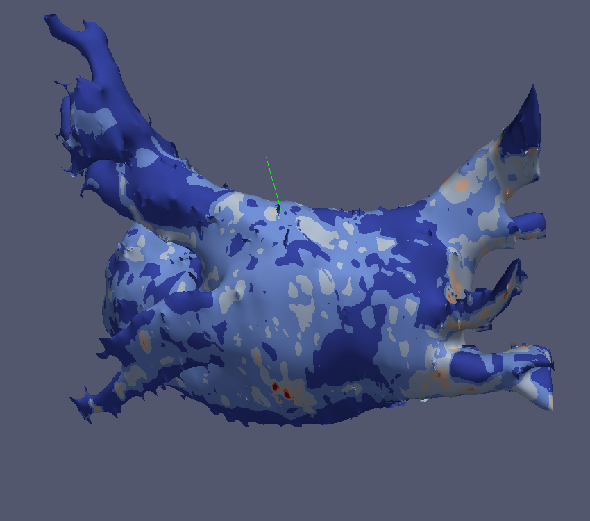
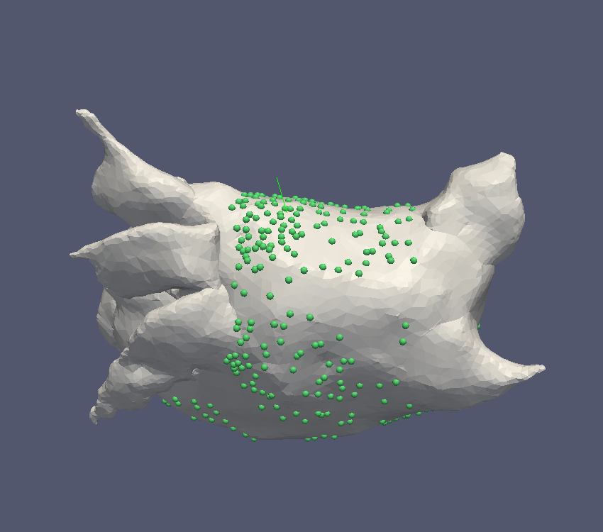
Motion Correction for Respiratory PET Images
(a) NCAT phantom with a lung lesion (blue).
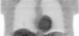
(b) Simulated PET image without motion correction. It is difficult to spot the lung lesion.
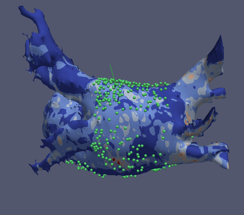
(c) After motion correction, it is much easier to see the lung lesion (red arrow).
Publications:
- W Bai and M Brady. Motion correction and attenuation correction for respiratory gated PET images. IEEE Transactions on Medical Imaging, 30(2):351-365, 2011.
- W Bai and M Brady. Regularized B-spline deformable registration for respiratory motion correction in PET images. Physics in Medicine and Biology, 54:2719-2736, 2009.
Microscopic Image Analysis
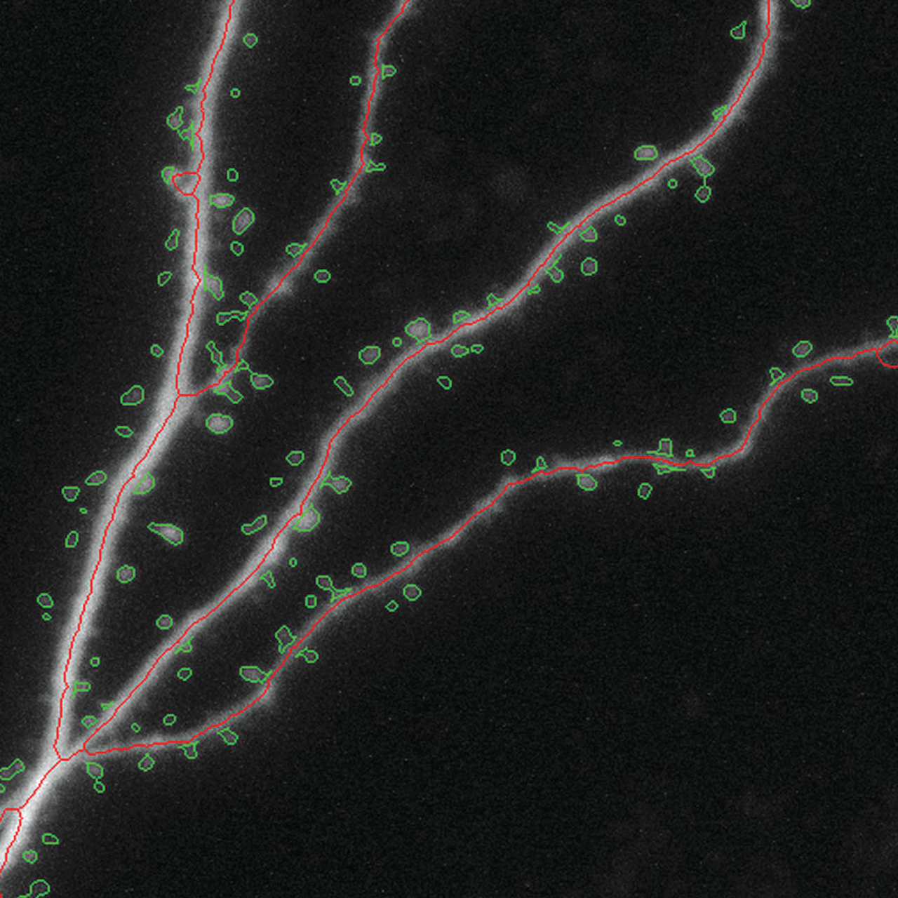
(a) Automatic segmentation of dendritic spines (green) and dendrite centrelines (red) in microscopic neuron images.
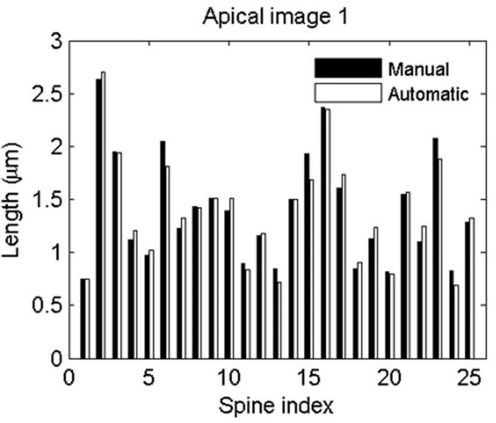
(b) The spine length measurements resulting from the automatic segmentation are very close to the manual measurements. The automatic method can greatly increase the efficiency of microscopic neuron image analysis.
Publications:
- W Bai, X Zhou, L Ji, J Cheng and STC Wong. Automatic dendritic spine analysis in two-photon laser scanning microscopy images. Cytometry A, 71A:818-826, 2007.