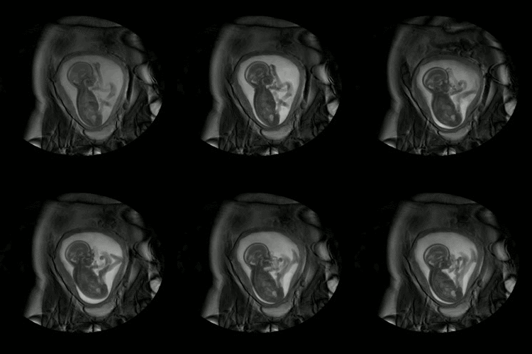
The aim is to develop and apply methods for quantitative Magnetic Resonance relaxometry and perfusion assessment in the fetal brain to create a comprehensive capability to measure key parameters in the presence of fetal motion. Conventional Magnetic Resonance Imaging (MRI) results in subject and scanner dependent signals rather than absolute measures. Relaxometry allows scanner a nd sequence independent unbiased signal maps to be obtained, which have the potential to provide a more objective readout. A complementary type of quantitative imaging focuses on functional measures rather than static tissue relaxation properties. One such measure focuses on tissue perfusion, which is rate of blood flow through unit mass of tissue within the micro-vasculature. MRI can be used to quantify perfusion, providing critical information that can be used both for scientific studies and for clinical care. Quantitative imaging requires the combination of multiple measurements at each tissue location, which is a major challenge in the presence of fetal motion.Having developed the methodology, two natural history studies will be conduct
ed – one in which T1 and T2 values are measured in fetal brain, and perhaps other organs, as a function of gestational age, and the other in which fetal brain perfusion is quantified again as a function of gestational age. The final result will provide techniques, to quantify physiological properties of the developing human, independent of scanner hardware and sequences.
If you are interested in doing a PhD in this area, consider applying for a PhD position in our EPSRC Centre for Doctoral Training in Medical Image Analysis!


