Research Highlights
A population-based phenome-wide association study of cardiac and aortic structure and function
Wenjia Bai et al. (2020) Nature Medicine volume 26, pages 1654–1662. DOI

“My research is at the interface between machine learning and medical imaging. I am interested in medical image segmentation, registration, reconstruction, classification and prediction problems, as well as their translation to clinical research and healthcare.”
Wenjia Bai
Differences in cardiac and aortic structure and function are associated with cardiovascular diseases and a wide range of other types of disease. Having developed tools for segmentation and image analysis of cardiac MR images using convolutional neural networks (for example, Bai et al. (2018a), Bai et al. (2018b)), these methods were used to develop an automated machine- learning-based analysis pipeline, which was used to analyse cardiovascular magnetic resonance images from a population-based study, the UK Biobank. In this paper, we report a comprehensive range of structural and functional phenotypes for the heart and aorta across 26,893 participants, and explore how these phenotypes vary according to sex, age and major cardiovascular risk factors.
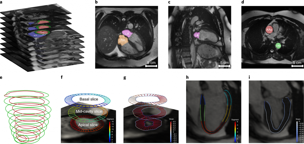
Fig. 1: Automated CMR image analysis pipeline.
Our automated pipeline evaluates comprehensive imaging phenotypes for the heart and aorta, including global phenotypes of the four cardiac chambers and two aortic sections: the left ventricle (LV), right ventricle (RV), left atrium (LA), right atrium (RA), ascending aorta (AAo) and descending aorta (DAo), as well as regional phenotypes of the LV myocardial-wall thickness and strain (Fig 1).
The derived imaging phenotypes from the 26,893 participants included in this study have been returned to UK Biobank to be made available for use by other researchers (https://biobank.ctsu.ox.ac.uk/crystal/dset.cgi?id=2383). A data visualization website was built (https://heartvis.doc.ic.ac.uk), which allows researchers to explore the associations between imaging phenotypes and non-imaging phenotypes. We have shared the image analysis pipeline (https://github.com/baiwenjia/ukbb_cardiac), which we anticipate will be a generally valuable resource for CMR image analysis.
We first characterized the associations of these phenotypes with sex and age in the population. Multiple-linear-regression models were built, using each imaging phenotype as the dependent variable and sex and age as independent variables. Overall, myocardial mass, cardiac-chamber volumes and aortic cross-sectional areas were greater for men than for women. Men consistently had lower cardiac-chamber ejection fractions than women did (Fig. 2).
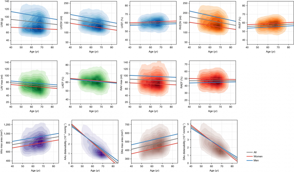
Fig. 2: Associations of selected imaging phenotypes with sex and age. Each graph displays a kernel density plot of an imaging phenotype plotted against age, as well as the linear-regression lines for the whole population (gray), for women (red) and for men (blue).
We explored the associations of cardiac and aortic imaging phenotypes with 12 categories of common diseases (Fig 3a), defined by self-reported diseases; for example, greater LVM was associated with a higher risk of hypertension and cardiac disease; greater cardiac-chamber volumes were associated with a higher risk of cardiac disease and with a lower risk of diabetes, asthma, chronic obstructive pulmonary disease (COPD) and bronchitis.
We also performed a phenome-wide association study (PheWAS) (Fig 3b) to explore the correlations between imaging phenotypes and 11 categories of non-imaging phenotypes of the participants. The non-imaging phenotypes include primary demographics, early-life factors, education and employment, diet, alcohol, smoking, physical activity, physical measures, self-reported medical conditions, mental health and cognitive function. We discovered a wide range of highly significant associations with lifestyle, early-life factors, mental health and cognitive function of the participants.
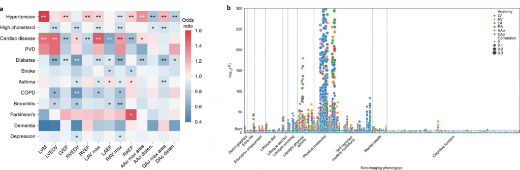
Fig. 3: (a) Associations of cardiac and aortic imaging phenotypes with common diseases; (b) Manhattan plot showing the P values (two-sided t-test) for correlations between imaging phenotypes and non-imaging phenotypes
On the basis of new observations in the PheWAS, we have reported significant associations of cardiac and aortic phenotypes with a wide range of participant phenotypes, including birth weight, mental health and cognitive-performance measures, illustrating the utility of generating these phenotypes even from a relatively healthy population when acquired at such a new scale. We investigated the effects of these non-imaging phenotypes on imaging phenotypes using an automated pipeline developed through the SmartHeart project.